Industrial Research and Technology Transfer Laboratory accredited by Emilia Romagna Region (DGR 1213/2007) with Deliberation n.GPG/2012/119 of February 6, 2012.
Industrial Research and Technology Transfer Laboratory accredited by Emilia Romagna Region (DGR 1213/2007) with Deliberation n.GPG/2012/119 of February 6, 2012.
The Flow Cytometry and Cell Sorting research group has acquired a great expertise in pre-clinical and biomedical research. In particular, the group is experienced (as certified by several publications on international scientific journals) in flow cytometry combined with molecular and cell biology techniques, applied in research projects principally concerning vascular disorders, stem cells and onco-haematology.
The Flow Cytometry and Cell Sorting service is equipped with the most advanced technology that allows many different research applications. In particular among the instruments, as detailed in the equipment section, the BD FACSAria IIIu cell sorter is especially powerful and versatile and it can be found in very few centers in Italy.
Examples of applications that the Flow Cytometry and Cell Sorting service can contribute to realize are:
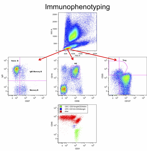
The service permits to: 1) obtain highly purified cell populations from various human and animal tissues by high throughput cell sorting; 2) phenotypically characterize isolated populations and/or any cell type circulating in human or laboratory animal peripheral blood; 3) monitor cell proliferation, survival and differentiation following in vitro and/or in vivo drug treatments. The Flow Cytometry and Cell Sorting service works in collaboration with the Biobank and Microscope services in characterizing cells of interest.
These applications concern both the in vitro evaluation of the effects produced by drugs, cytokines or other compounds and also the research aimed at broadening the clinical understanding of various diseases.
High performance cytometer and cell sorter, provided with 4 laser sources (blue - 488 nm, red - 633 nm, violet - 405 nm, near UV - 375 nm), it can detect up to 13 parameters, 11 of which can be fluorescence parameters.
The number of installed lasers, in combination with the revolutionary optical bench, greatly increases the number of fluorescences that the instrument is capable of analyzing at one time and consequently the range of usable fluorochromes.
The higher performance of the excitation and fluorescence detection system, the electronics and the FACSDiva software 9, together with the easiness of use and the quickness of instrument setup, due to the fixed alignment technology, that also ensures a very high reproducibility, make the FACSAria IIIu the ideal cell sorter for modern biomedical research laboratories.
| Lasers (exitation) |
PMTs | BP Filters (emission) |
Fluorochromes (examples) |
| *Note: nearUV and Violet lasers cannot be used at the same time. | |||
| Blue 488 nm |
A FL1 | 780/80 | PE-Cy7, PE-Vio770, Qdot800 |
| B FL2 | 698/40 | PerCP, PerCP-Cy5.5, PerCP-Vio700, PerCP-eFluor710, 7-AAD, PI | |
| C FL3 | 616/23 | PE-TexasRed | |
| D FL4 | 585/42 | PE | |
| E FL5 | 530/30 | FITC, Alexa 488, GFP, Sytox Green, CFSE, DCFDA | |
| F SSC | 488/10 | SSC | |
| Red 633 nm |
A FL6 | 780/60 | APC-H7, APC-Vio770, Live/DEAD Near-IR |
| B FL7 | 730/45 | AF700 | |
| C FL8 | 660/20 | APC | |
| Violet 405 nm* |
A FL9 | 780/60 | BV750, BV785 |
| B FL10 | 530/30 | V500, VioBlue, AmCyan, BV510, Pacific Orange, LIVE/DEAD Aqua | |
| C FL11 | 450/40 | V450, VioBlue, Pacific Blue, CellTracker Violet, Zombie violet, AF405 | |
| Near UV 375 nm* |
A FL9 | 780/60 | BUV737, BUV805 |
| B FL10 | 530/30 | Indo-1 (Blue) | |
| C FL11 | 450/40 | Hoechst Blue, DAPI | |
Bench flow cytometer with two light sources spatially separate: an air-cooled argon laser with an emission at 488 nm and a red diode laser (Visible Red Diode Laser) with an emission at 635 nm. The above described optical configuration allow the analysis of up to 6 parameters, 4 of which can be fluorescence parameters, and a great flexibility in the choice of fluorochromes.
Specifically designed to support a wide range of applications, the FACScalibur is characterized by an high level of automation (instrument control software and digital fluidics control) to guarantee the higher reproducibility of the results.
Our facility also makes use of the FlowJo software for data analysis (licence dongles available for signing out for short times). This powerful flow cytometry analysis program is compatible with data acquired from any flow cytometry instruments and capable of executing complex analysis, making the process of analisis of multiparametric acquisitions more efficient, easier and faster.
| Lasers (exitation) |
PMTs | BP Filters (emission) |
Fluorochromes (examples) |
| Blue 488 nm |
A FL1 | 530/30 | FITC, Alexa 488, GFP, Sytox Green, CFSE, DCFDA |
| B FL2 | 585/42 | PE | |
| C FL3 | 670 LP | PerCP, PerCP-Cy5.5, PerCP-Vio700, PerCP-eFluor710, 7-AAD, PI | |
| Red 633 nm |
D FL4 | 661/16 | APC |
Flow Cytometry and Cell Sorting facility
Area Polo Chimico Biomedico ‘CUBO’ – third floor
Via Fossato di Mortara 70 - 44121 Ferrara
+39 0532 455936/718/849
+39 0532 455718
Prof. Elisabetta Melloni
elisabetta.melloni@unife.it
+39 0532 455936
Dott. Fabio Casciano
fabio.casciano@unife.it
+39 0532 455718
Dr Fabio Casciano
Dr Elisabetta Melloni
Our staff is qualified to i) receive and, if needed, process/stain the samples, ii) characterize and/or isolate cells of interest, iii) analyze results, and iv) give a quality control of them.
| Casciano F, Diani M, Altomare A, Granucci F, Secchiero P, Banfi G, Reali E (2020). CCR4+ Skin-Tropic Phenotype as a Feature of Central Memory CD8+ T Cells in Healthy Subjects and Psoriasis Patients. Front Immunol; 11:529. doi: 10.3389/fimmu.2020.00529. PubMed |
| Diani M, Casciano F, Marongiu L, Longhi M, Altomare A, Pigatto PD, Secchiero P, Gambari R, Banfi G, Manfredi AA, Altomare G, Granucci F, Reali E (2019). Increased frequency of activated CD8+ T cell effectors in patients with psoriatic arthritis. Sci Rep; 9(1):10870. doi: 10.1038/s41598-019-47310-5. doi: 10.1038/s41598-019-47310-5. PubMed |
| Chiriaco M, Casciano F, Di Matteo G, Gentner B, Claps A, Di Cesare S, Cotugno N, D'Argenio P, Rossi P, Aiuti A, Finocchi A (2018). Impaired X-CGD T cell compartment is gp91phox-NADPH oxidase independent. Clin Immunol; 193:52-59. doi: 10.1016/j.clim.2018.01.010. PubMed |
| D'Abundo L, Callegari E, Bresin A, Chillemi A, Elamin BK, Guerriero P, Huang X, Saccenti E, Hussein E, Casciano F, Secchiero P, Zauli G, Calin GA, Russo G, Lee LJ, Croce CM, Marcucci G, Sabbioni S, Malavasi F, Negrini M (2017). Anti-leukemic activity of microRNA-26a in a chronic lymphocytic leukemia mouse model. Oncogene; 36(47):6617-6626. doi: 10.1038/onc.2017.269. PubMed |
| Secchiero P, Voltan R, Rimondi E, Melloni E, Athanasakis E, Tisato V, Gallo S, Rigolin GM, Zauli G (2017). The γ-secretase inhibitors enhance the anti-leukemic activity of Ibrutinib in B-CLL cells. Oncotarget; 8(35):59235-59245. doi:10.18632/oncotarget.19494. PubMed |
| Feriotto G, Calza R, Bergamini CM, Griffin M, Wang Z, Beninati S, Ferretti V, Marzola E, Guerrini R, Pagnoni A, Cavazzini A, Casciano F, Mischiati C (2016). Involvement of cell surface TG2 in the aggregation of K562 cells triggered by gluten. Amino Acids 49(3):551-565. doi: 10.1007/s00726-016-2339-4. PubMed |
| Voltan R, Rimondi E, Melloni E, Gilli P, Bertolasi V, Casciano F, Rigolin GM, Zauli G, Secchiero P (2016). Metformin combined with sodium dichloroacetate promotes B leukemic cell death by suppressing anti-apoptotic protein Mcl-1. Oncotarget 7(14):18965-77. doi: 10.18632/oncotarget.7879. PubMed |
| Trapella C, Voltan R, Melloni E, Tisato V, Celeghini C, Bianco S, Fantinati A, Salvadori S, Guerrini R, Secchiero P, Zauli G (2016). Design, Synthesis, and Biological Characterization of Novel Mitochondria Targeted Dichloroacetate-Loaded Compounds with Antileukemic Activity. J Med Chem. 59(1):147-56. doi:10.1021/acs.jmedchem.5b01165. PubMed |
| Agnoletto C, Melloni E, Casciano F, Rigolin GM, Rimondi E, Celeghini C, Brunelli L, Cuneo A, Secchiero P, Zauli G (2014). Sodium dichloroacetate exhibits anti-leukemic activity in B-chronic lymphocytic leukemia (B-CLL) and synergizes with the p53 activator Nutlin-3. Oncotarget 5:4347-60. doi:10.18632/oncotarget.2018. PubMed |
| Athanasakis E, Melloni E, Gian Matteo Rigolin GM, Agnoletto C, Voltan R, Vozzi D, Piscianz E, Segat L, Dal Monego S, Cuneo A, Secchiero P, Zauli G (2014). The p53 transcriptional pathway is preserved in ATMmutated and NOTCH1mutated chronic lymphocytic leukemias. Oncotarget; 5(24):12635-45. doi:10.18632/oncotarget.2211. PubMed |
| Code no. | Brief description of the services we offer | Price (€) |
| Preliminary consultation on formulation of experimental design; during the consultation an estimate of the overall cost of the analyses can be provided. The Service offers consultations for specific projects, and estimates of the overall cost are provided free of charge according to the needs of the client. Please contact the representative for special requirements not shown in the price list, since the packages offered can be tailored to the customer’s requirements. For routine analysis prices can be arranged (subscriptions may be possible). |
No cost | |
| CF-01 | Immunophenotyping of 1 surface antigen. The procedure consist of antibody labelling, data acquisition and analysis. |
60/hour |
| CF-02 | Immunophenotyping of 2-4 antigens through detection with specific fluorophore-conjugated antibodies. The procedure consist of compensation matrix, antibody labelling, data acquisition and analysis. |
70/hour |
| CF-03 | Apoptosis quantification (monoparametric with PI). The procedure consist of staining, data acquisition and analysis. |
50/hour |
| CF-04 | Apoptosis quantification (biparametric with AnnexinV/PI or 7-AAD). The procedure consist of antibody labelling and staining, data acquisition and analysis. |
70/hour |
| CF-05 | Cell cycle analysis (monoparametric with PI). The procedure consist of staining, data acquisition and analysis. |
50/hour |
| CF-06 | Cell cycle analysis (biparametric with BrdU-FITC/PI). The procedure consist of antibody labelling and staining, data acquisition and analysis. |
70/hour |
| CS-01 | Immunophenotyping of 5 or more surface antigens. The procedure consist of antibody labelling, compensation matrix data acquisition and analysis. |
80/hour |
| CS-02 | Monoparametric sorting (cells isolated according to the presence of GFP or of 1 surface antigen). The procedure consist of antibody labelling, set-up using AccuDrop test, data acquisition, sorting, check of the sorting purity. |
85/hour |
| CS-03 | Multiparametric sorting (cells isolated according to the presence of 2 or more surface antigens). The procedure consist of antibody labelling, set-up using AccuDrop test, compensation matrix, data acquisition, sorting, check of the sorting purity. |
110/hour |
| CFCS-01 | Multiparametric analysis of the expression of cytokines, transcription factors and phospho-proteins using specific kits, also in combination with surface immunophenotyping. | |
| CFCS-02 | Expansion of cell lines and possible treatment with exogenous substances aimed at supply specific Cytometry and Cell Sorting services. | To agree with the customer |
| CFCS-03 | Consultancy/ technical-practical courses/ other services | To define depending of the customer’s requirements |
Some of the services/products offered may be provided in collaboration/integration with other services of the LTTA Laboratory.
Each service includes the supply of images and/or files for scientific publication.
Prices are shown without VAT and are subjected to change according to the cost of consumables (every 12 months).
LTTA c/o CUBO
Via Fossato di Mortara, 70
44121 Ferrara
Main Entrance • Piazzale Eliporto

Web design and
site maintenance
Azuleon sas
© 2011-2024