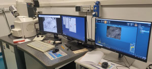
The Electron Microscopy Centre can provide technical expertise and scientific equipment for studying ultrastructures, whether for life sciences or materials science. Indeed, our staff have solid experience in the scientific sector, particularly regarding the use and application of electron microscopy (transmission, scanning, atomic force and X-ray microanalysis), and their work includes preparing samples, viewing and interpreting images. The use of the electron microscope, allows to achieve image resolution several orders of magnitude greater than with an optical microscope, and, for example, to view thin sections of tissue enlarged so that it is possible to study the subcellular structures (intracytoplasmic organelles, cytoskeleton, etc) and macromolecular components of the tissue stroma (collagen, protocollagen, etc).
The Electron Microscopy Centre collaborates with researchers from the university, hospitals, public and private bodies, industry and individual professionals, providing access to state-of-the-art scientific equipment and highly specialized staff to provide the specific technical expertise for the preparation and observation of samples using electron and atomic force microscopy.
The applications cover various fields of interest in a wide range of disciplines, for example: animal and plant biology, materials science, rock and mineral science, chemistry, geochemistry, micropalaeontology, cultural assets and environmental problems, medical and clinical science.
We use the following basic techniques in our work at the Centre.
Scanning electron microscopy (SEM): informations can be obtained about the morphology and surface topography of very large areas (one mm side length) down to submicrometer scale. Microanalysis with SEM can identify chemical elements present in selected areas and in particles larger than one micron. The energy dispersive spectrometer (EDS) provide quantitative analysis of elements with an atomic number lower than 10, as well as the compositional maps. In addition to conductive materials, non-conductor (insulating) samples can also be viewed, without the use of a metal film as required for conventional SEM: for example, items encased in resin, ceramics, card, polymers, ambient dust, plant fragments, and fixed dehydrated tissue of animal or human origin.
Transmission electron microscopy (TEM): the ultrastructure of biological preparations and the morphology of nanoparticles can be observed, and defective areas in crystalline materials identified. It is possible to observe ultrathin layers of biological preparations, dust, deposited films and thin metal fragments.
Atomic force microscopy (AFM): surface topography down to nanometric level, the dimensions of details even in vertical direction to the plane of the sample (section analysis), roughness, the Fourier spectrum, the presence of magnetic domains of submicrometric dimension (in MFM) in combination with the topography of the area scanned can be determined. It is possible to observe deposited films, metals, polymers, plastic materials, ceramic products, magnetic tapes, dried histological preparations and plant fragments.
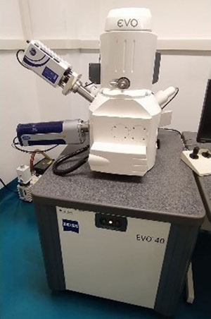
Zeiss EVO 40 Scanner electron microscope (SEM):

Microscopio elettronico a scansione: SEM EVO Zeiss

Microscopio elettronico a scansione: SEM EVO Zeiss detector EBSD
Transmission electron microscope, Zeiss EM 910 (TEM):
Microscopio elettronico a scansione in Field Emission, (FEG SEM) Zeiss Gemini 460:
Microscopio elettronico a trasmissione (TEM), ThermoFisher TalosTM L120C G2:
Microscopio Iperspettrale Cytoviva:
Preparation equipment:
Electron Microscopy at the Electron Microscopy Centre of the University of Ferrara, Polo Chimico Biomedico
Via Luigi Borsari 46 - 44121 Ferrara
+39 0532 455 940/845/725
+39 0532 455064
Prof. Luca Maria Neri
luca.neri@unife.it
+39 0532 455940
Dr Erika Rimondi (Technical Coordinator CME)
erika.rimondi@unife.it
+39 0532 455845
Dr Paola Boldrini
Dr Cinzia Brenna
Dr Edi Simoni
The work of our staff involves: i) analysing problems posed by clients; ii) formulating suitable strategies for solving the problems; iii) receiving samples; iv) preparing samples for observations under the microscope; v) providing support for the observation, preparation analysis and interpretation of data.
| Sfriso AA, Mistri M, Munari C, Moro I, Wahsha M, Sfriso A, Juhmani AS. Environ Int. (2019) Hazardous effects of silver nanoparticles for primary producers in transitional water systems: The case of the seaweed Ulva rigida C. Agardh. 131:104942. doi: 10.1016/j.envint.2019.104942 |
| Pezzi M, Scapoli C, Mamolini E, Leis M, Bonacci T, Whitmore D, Krčmar S, Furini M, Giannerini S, Chicca M, Cultrera R, Faucheux MJ (2018) Ultrastructural characterization of sensilla and microtrichia on the antenna of female Haematopota pandazisi (Diptera: Tabanidae). Parasitol Res. 117(4):959-70. doi: 10.1007/s00436-018-5760-7. PubMed |
| Caselli E, Pancaldi S, Baldisserotto C, Petrucci F, Impallaria A, Volpe L, D'Accolti M, Soffritti I, Coccagna M, Sassu G, Bevilacqua F, Volta A, Bisi M, Lanzoni L, Mazzacane S (2018) Characterization of biodegradation in a 17th century easel painting and potential for a biological approach. PLoS One. 13:e0207630. doi: 10.1371/journal.pone.0207630. PubMed |
| Zamboni P, Giaquinta A, Rimondi E, Pedriali M, Scanziani E, Riccaboni P, Veroux M, Secchiero P, Veroux P (2018) A novel endovenous scaffold for the treatment of chronic venous obstruction in a porcine model: Histological and ultrastructural assessment. Phlebology. 18:268355518805686. doi: 10.1177/0268355518805686. PubMed |
| Munari C, Bocchi N, Parrella P, Granata T, Moruzzi L, Massara F, De Donati M, Mistri M (2017) The occurrence of two morphologically similar Chaetozone (Annelida: Polychaeta: Cirratulidae) species from the Italian seas: Chaetozone corona Berkeley & Berkeley, 1941 and C. carpenteri McIntosh, 1911. The European Zoological Journal. 84(1): 541-553. doi.org/10.1080/24750263.2017.1393843 |
| Di Lecce D, Hu T, Hassoun J (2017) Electrochemical features of LiMnPO4 olivine prepared by sol-gel pathway. Journal of Alloys and Compounds. 93:730-37. doi: 10.1016/j.jallcom.2016.09.193 |
| Dezfuli BS, DePasquale JA, Castaldelli G, Giari L, Bosi G (2017) A fish model for the study of the relationship between neuroendocrine and immune cells in the intestinal epithelium: Silurus glanis infected with a tapeworm. Fish Shellfish Immunol. 64:243-250. doi: 10.1016/j.fsi.2017.03.033. PubMed |
| Bosi G, Giari L, DePasquale JA, Carosi A, Lorenzoni M, Dezfuli BS (2017) Protective responses of intestinal mucous cells in a range of fish-helminth systems. J Fish Dis. 40(8):1001-1014. doi: 10.1111/jfd.12576. PubMed |
| Lombardo L, Martini M, Cervinara F, Spedicato GA, Oliverio T, Siciliani G (2017) Comparative SEM analysis of nine F22 aligner cleaning strategies. Prog Orthod. 18(1):26. doi: 10.1186/s40510-017-0178-9. PubMed |
| Pezzi M, Leis M, Chicca M, Falabella P, Salvia R, Scala A, Whitmore D (2017) Morphology of the Antenna of Hermetia illucens (Diptera: Stratiomyidae): An Ultrastructural Investigation. J Med Entomol. 54:925-33. doi: 10.1093/jme/tjx055. PubMed |
| Code no. | Brief description of the services we offer | Price (€ per hour) |
| Transmission electron microscopy - TEM | ||
| TEM-001 | Fixation | 122.90 |
| TEM-002 | Embedding | 69.60 |
| Ultramicrotomy (1 Block) | ||
| TEM-003 | A. semi-thin sections | 8.20 |
| TEM-004 | B. stained ultrathin sections | 46.10 |
| TEM-005 | Observation (1 hour) | 122.90/hour |
| Scanning electron microscopy - SEM | ||
| SEM-001 | Fixation | 122.90 |
| SEM-002 | Critical point dryer | 28.70 |
| SEM-003 | Metallization with gold, up to 7 samples | 61.40 |
| SEM-004 | Metallization with graphite, up to 7 samples | 15.40 |
| SEM-005 | Observation | 122.90/hour |
| SEM-006 | X-ray microanalysis | 122.90/hour |
Alcune prestazioni/prodotti offerti possono usufruire della collaborazione/integrazione con altri servizi del Laboratorio LTTA.
Il centro di microscopia elettronica offre possibilità di tirocinio curricolare per studenti iscritti a corsi di laurea triennale e magistrale.
The digital and confocal microscopy service is a centre dedicated to innovative technology in cellular imaging. The remarkable technological progress seen in recent years in research instruments and image analysis has meant higher resolution and contrast can be obtained even from complex samples, opening up significant new perspectives in the field of experimental biology. At same time, the development of specific synthetic engineered probes to mark and dynamically monitor cell structures or physiological parameters has enabled researchers to accurately distinguish different subcellular structures in a single experiment.
The advanced microscopy centre acts as a point of access to highly competitive, sophisticated instruments, image acquisition methods and analytical methods. Live imaging has become an essential instrument in many research laboratories and a basic methodology in many medical disciplines, in order to study cell architecture and analyse in real time dynamic molecular processes in living cells, helping to understand how cells and tissues function. It also enables high throughput assays in biological processes to be performed, thereby extending the field of application and interest to pharmaceutical companies.
Confocal microscopy offers several advantages, such as control of depth of field, a notable improvement in image quality, and the possibility of performing serial optical sections, obtaining high resolution 3D images and offering the possibility of analysing dynamic intracellular events. Live imaging requires very short acquisition times to capture extremely fast events occurring in cells. For this purpose the microscopy centre has an extended field confocal system (Nikon LiveScan Swept Field Confocal microscope), which is ideal for imaging very fast intracellular events, such as, for example, dynamics of calcium ions events which are one of the laboratory’s areas of expertise. We also have an Olympus screening platform, designed for totally automated image acquisition and data analysis optimised for viewing very high speed processes in live cells. These systems are the perfect instruments for developing assays and performing high content screening, combining modularity and flexibility in a microscope with automation, reproducibility and reliability of results. The technology in our laboratory, combined with our decades of experience in microscopy, the availability of recombinant probes and our expertise in developing the most suitable protocols, allows us to meet the needs of the scientific community and offer functional screening to track dynamic changes over time in hundreds of thousands of living cells.
The department is moreover actively involved in the development of a new software for high-resolution imaging problems, able to take advantage of both multi-processor and massively parallel computation (GPU) architectures. These software will be tested both on clusters at University of Ferrara and on European level facilities, as the BG/Q architecture of recent installation at the CINECA consortium.
Also, the plate reader XF96 Extracellular Flux Analyzer, Seahorse Bioscience, which has been recently acquired in the laboratory, permits the measurements in real-time of metabolic activities in live cells, quantifying simultaneously both mitochondrial respiration and glycolysis.
Finally, many researchers can usefully benefit from access to this state-of-the-art technology, particularly now as the post-genome era in biomedical research begins.
The advanced confocal microscopy centre at the LTTA laboratory is a highly competitive service providing technical expertise and sophisticated, innovative instruments. Its field of applicability ranges from basic biomedical research to more specific services, such as screening analysis of molecular activity of interest in the field of pharmaceuticals. The centre has many state-of-the-art instruments, such as the Olympus xcellence platform/ScanˆR for live cell imaging and high content screening, and a confocal microscopy system from Nikon for studying very rapid, dynamic intracellular events.
Recently we have acquired the XF96 Extracellular Flux Analyzer, Seahorse Bioscience, a plate reader for studies of cellular metabolic activities.
Live monitoring of cell behaviour, using specific probes, enables a very wide range of analyses to be performed, thus extending the field of application from basic research to more specific studies, such as the screening of pharmacologically active molecules by monitoring their proliferation, survival and cellular differentiation following in vitro and/or in vivo drug treatment; specifically:
Scan^R workstation
An automated station for digital imaging of cytometric parameters in living cells. The automatic recognition of objects enables the use of high content/throughput protocols to be made for principal cellular events (apoptosis, necrosis, autophagy/mitophagy, cell cycles, morphological changes, protein expression, automated analysis for FISH, and protein localisation and translocation.
Xcellence workstation
Multiple wavelength high resolution fluorescence microscopy system. The system’s high resolution enables 2D/3D analysis of intracellular structures and organisation to be made (i.e. mitochondria, endoplasmic reticulum, and cytoskeleton). Moreover, the multiple wavelengths and versatility of the system enable live measurement of ratiometric probes such as fura or pericam to be performed.
Live Scan Swept Field Confocal
High-speed confocal fluorescence microscopy system for in vivo analysis of multiple cell parameters. The system enables simultaneous measurement of cell parameters such as calcium concentration (cytoplasmic/mitochondrial), protein translocation, reorganisation of cell structure, and vesicular generation, loss and fusion. Three excitation lines are fitted, based on solid-state lasers (488nm, 561nm, 635nm), and two different emission filter sets (488/561, 488/643). This system enables two different fluorophores to be visualised at the same time.
Seahorse XF96 Extracellular Flux Analyzer
The Seahorse XF96 Extracellular Flux Analyzer simultaneously measures mitochondrial respiration and glycolysis in cells in real-time. This instrument provides the most physiologically relevant in vitro measurement for your cell metabolism research in primary cells and tumor cell lines, either adherent or suspension cells, as well as islets and isolated mitochondria; it is based on calibrated plates with fluorescent compounds for the simultaneous measurement of oxygen levels and pH in the medium. By measuring the key parameters of cell metabolism, it provides functional data that enables a greater understanding of cancer, aging, and metabolic, cardiovascular, and neurodegenerative diseases.
Advanced Digital and Confocal Microscopy facility
Laboratorio per le Tecnologie delle Terapie Avanzate (LTTA)
Area Polo Chimico Biomedico ‘CUBO’ – second floor
Via Fossato di Mortara 70 - 44121 Ferrara
+39 0532 455806
+39 0532 455351
Prof. Paolo Pinton
pnp@unife.it
+39 0532 455802
Dr Sonia Missiroli
sonia.missiroli@unife.it
+39 0532 455806
Dr Carlotta Giorgi
Dr Massimo Bonora
Dr Sonia Missiroli
The work of our staff involves i) analysis of problems posed by clients, ii) receiving and preparing samples, iii) designing and performing microsopic assays, iv) analysis of the results, v) sending all the information to the user for correct interpretation.
| Kuchay S, Giorgi C, Simoneschi D, Pagan J, Missiroli S, Saraf A, Florens L, Washburn MP, Collazo-Lorduy A, Castillo-Martin M, Cordon-Cardo C, Sebti SM, Pinton P, Pagano M (2017) PTEN counteracts FBXL2 to promote IP3R3- and Ca2+-mediated apoptosis limiting tumour growth. Nature 546:554-558. PubMed |
| Bonora M, Morganti C, Morciano G, Pedriali G, Lebiedzinska-Arciszewska M, Aquila G, Giorgi C, Rizzo P, Campo G, Ferrari R, Kroemer G, Wieckowski MR, Galluzzi L, Pinton P (2017) Mitochondrial permeability transition involves dissociation of F1FO ATP synthase dimers and C-ring conformation. EMBO Rep. 18:1077-1089. PubMed |
| Bonora M, Morganti C, Morciano G, Giorgi C, Wieckowski MR, Pinton P (2016) Comprehensive analysis of mitochondrial permeability transition pore activity in living cells using fluorescence-imaging-based techniques. Nat Protoc 11:1067-80. PubMed |
| Missiroli S, Bonora M, Patergnani S, Poletti F, Perrone M, Gafà R, Magri E, Raimondi A, Lanza G, Tacchetti C, Kroemer G, Pandolfi PP, Pinton P, Giorgi C (2016) PML at Mitochondria-Associated Membranes Is Critical for the Repression of Autophagy and Cancer Development. Cell Rep 16:2415-27. PubMed |
| Rimessi A, Bezzerri V, Patergnani S, Marchi S, Cabrini G, Pinton P (2015) Mitochondrial Ca2+-dependent NLRP3 activation exacerbates the Pseudomonas aeruginosa-driven inflammatory response in cystic fibrosis. Nat Commun 6:6201. PubMed |
| Giorgi C, Bonora M, Missiroli S, Poletti F, Ramirez FG, Morciano G, Morganti C, Pandolfi PP, Mammano F, Pinton P (2015) Intravital imaging reveals p53-dependent cancer cell death induced by phototherapy via calcium signaling. Oncotarget 6:1435-45. PubMed |
| Bonora M, De Marchi E, Patergnani S, Suski JM, Celsi F, Bononi A, Giorgi C, Marchi S, Rimessi A, Duszyński J, Pozzan T, Wieckowski MR, Pinton P (2014) Tumor necrosis factor-α impairs oligodendroglial differentiation through a mitochondria-dependent process. Cell Death Differ. 21:1198-208. PubMed |
| Rimessi A, Marchi S, Patergnani S, Pinton P (2014) H-Ras-driven tumoral maintenance is sustained through caveolin-1-dependent alterations in calcium signaling. Oncogene 33:2329-40. PubMed |
| Marchi S, Patergnani S, Pinton P (2014) The endoplasmic reticulum-mitochondria connection: one touch, multiple functions. Biochim. Biophys. Acta 1837:461-9. PubMed |
| Marchi S, Lupini L, Patergnani S, Rimessi A, Missiroli S, Bonora M, Bononi A, Corrà F, Giorgi C, De Marchi E, Poletti F, Gafà R, Lanza G, Negrini M, Rizzuto R, Pinton P (2013) Downregulation of the mitochondrial calcium uniporter by cancer-related miR-25. Curr. Biol. 23:58-63. PubMed |
| Bonora M, Bononi A, De Marchi E, Giorgi C, Lebiedzinska M, Marchi S, Patergnani S, Rimessi A, Suski JM, Wojtala A, Wieckowski MR, Kroemer G, Galluzzi L, Pinton P (2013) Role of the c subunit of the FO ATP synthase in mitochondrial permeability transition. Cell Cycle 12:674-83. PubMed |
| Chiabrando D, Marro S, Mercurio S, Giorgi C, Petrillo S, Vinchi F, Fiorito V, Fagoonee S, Camporeale A, Turco E, Merlo GR, Silengo L, Altruda F, Pinton P, Tolosano E (2012) The mitochondrial heme exporter FLVCR1b mediates erythroid differentiation. J. Clin. Invest. 122:4569-79. PubMed |
| Garcia-Cao I, Song MS, Hobbs RM, Laurent G, Giorgi C, de Boer VC, Anastasiou D, Ito K, Sasaki AT, Rameh L, Carracedo A, Vander Heiden MG, Cantley LC, Pinton P, Haigis MC, Pandolfi PP (2012) Systemic elevation of PTEN induces a tumor-suppressive metabolic state. Cell 149:49-62. PubMed |
| Pinton P, Giorgi C, Pandolfi PP (2011) The role of PML in the control of apoptotic cell fate: a new key player at ER-mitochondria sites. Cell Death Differ. 18:1450-6. PubMed |
| Giorgi C, Ito K, Lin HK, Santangelo C, Wieckowski MR, Lebiedzinska M, Bononi A, Bonora M, Duszynski J, Bernardi R, Rizzuto R, Tacchetti C, Pinton P, Pandolfi PP (2010) PML regulates apoptosis at endoplasmic reticulum by modulating calcium release. Science 330:1247-51. PubMed |
| Demaria M, Giorgi C, Lebiedzinska M, Esposito G, D'Angeli L, Bartoli A, Gough DJ, Turkson J, Levy DE, Watson CJ, Wieckowski MR, Provero P, Pinton P, Poli V (2010) A STAT3-mediated metabolic switch is involved in tumour transformation and STAT3 addiction. Aging (Albany NY) 2:823-42. PubMed |
| Goitre L, Balzac F, Degani S, Degan P, Marchi S, Pinton P, Retta SF (2010) KRIT1 regulates the homeostasis of intracellular reactive oxygen species. PLoS ONE 5:e11786. PubMed |
| Pavan C, Vindigni V, Michelotto L, Rimessi A, Abatangelo G, Cortivo R, Pinton P, Zavan B (2010) Weight gain related to treatment with atypical antipsychotics is due to activation of PKC-β. Pharmacogenomics J. 10:408-17. PubMed |
| Rimessi A, Marchi S, Fotino C, Romagnoli A, Huebner K, Croce CM, Pinton P, Rizzuto R (2009) Intramitochondrial calcium regulation by the FHIT gene product sensitizes to apoptosis. Proc. Natl. Acad. Sci. U.S.A. 106:12753-8. PubMed |
| Aguiari P, Leo S, Zavan B, Vindigni V, Rimessi A, Bianchi K, Franzin C, Cortivo R, Rossato M, Vettor R, Abatangelo G, Pozzan T, Pinton P, Rizzuto R (2008) High glucose induces adipogenic differentiation of muscle-derived stem cells. Proc. Natl. Acad. Sci. U.S.A. 105:1226-31. PubMed |
| Rimessi A, Rizzuto R, Pinton P (2007) Differential recruitment of PKC isoforms in HeLa cells during redox stress. Cell Stress Chaperones 12:291-8. PubMed |
| Pinton P, Rimessi A, Marchi S, Orsini F, Migliaccio E, Giorgio M, Contursi C, Minucci S, Mantovani F, Wieckowski MR, Del Sal G, Pelicci PG, Rizzuto R (2007) Protein kinase C beta and prolyl isomerase 1 regulate mitochondrial effects of the life-span determinant p66Shc. Science 315:659-63. PubMed |
| Pinton P, Leo S, Wieckowski MR, Di Benedetto G, Rizzuto R (2004) Long-term modulation of mitochondrial Ca2+ signals by protein kinase C isozymes. J. Cell Biol. 165:223-32. PubMed |
| Rapizzi E, Pinton P, Szabadkai G, Wieckowski MR, Vandecasteele G, Baird G, Tuft RA, Fogarty KE, Rizzuto R (2002) Recombinant expression of the voltage-dependent anion channel enhances the transfer of Ca2+ microdomains to mitochondria. J. Cell Biol. 159:613-24. PubMed |
| Rizzuto R, Pinton P, Carrington W, Fay FS, Fogarty KE, Lifshitz LM, Tuft RA, Pozzan T (1998) Close contacts with the endoplasmic reticulum as determinants of mitochondrial Ca2+ responses. Science 280:1763-6. PubMed |
The optical microscopy centre regularly organises courses on Digital and Confocal Microscopy open to anyone who is interested in learning the basic elements necessary for using confocal microscopy in the field of biology. The courses include theory classes and practical sessions using the equipment, in order to learn the methods and procedures for acquiring high quality informative images. The closing dates for registration will be shown in the annual calendar. At the end of the course those attending will be presented with a certificate of participation.
| Code no. | Brief description of the services we offer | Price (€ per hour) |
| Preliminary consultation on formulation of experimental design and/or sample preparation; during the consultation a quote can be prepared on the overall cost of the analyses required. The Service offers consultations for specific projects, and quotes are provided free of charge according to the needs of the user. Please contact us for individual requirements not shown in the price list, since the packages offered can be tailored to requirements outside the standard service. For routine analysis prices can be agreed (subscription may be possible). |
0.00 | |
| Nikon Confocal | ||
| MC-01 | Sample preparation | 100.00 |
| Sample observation | ||
| MC-02 | - Assistance from staff | 120.00 |
| MC-03 | - Acquisition | 130.00 |
| MC-04 | Data analysis | 140.00 |
| MC-05 | Consumables | according to materials used |
| Xcellence | ||
| MC-06 | Sample preparation | 100.00 |
| Sample observation | ||
| MC-07 | - Assistance from staff | 120.00 |
| MC-08 | - Acquisition | 30.00 |
| MC-09 | Data analysis | 140.00 |
| MC-10 | Consumables | according to materials used |
| Scan^R HTM | ||
| MC-11 | Sample preparation | 100.00 |
| Sample observation | ||
| MC-12 | - Assistance from staff | 120.00 |
| MC-13 | - Acquisition | 70.00 |
| MC-14 | Data analysis | 140.00 |
| MC-15 | Consumables | according to materials used |
| Theory and Practical Courses | ||
| MC-16 | Theory Course | 1000.00 |
| MC-17 | Practical Course (€ /hour / person) (up to four persons per group). No further expense is required for use of the equipment. |
120.00 |
| Independent Use (Only if authorized) | ||
| Sample observation | ||
| MC-18 | -Nikon | 120.00 |
| MC-19 | -Xcellence | 30.00 |
| MC-20 | -Scan^R | 130.00 |
| MC-21 | Use of Workstation for data analysis | 70.00 |
| MC-22 | Consumables | according to materials used |
| XF96 Extracellular Flux Analyzer | ||
| MC-23 | 1Assistance from staff | 120.00€/hour |
| MC-24 | 2Mito stress assay (mito stress kit, cell culture microplate, XF assay medium, sensor cartridge, XF calibrant) | 270.00€/experiment |
| MC-25 | 2Glycolysis stress assay (glycolysis stress kit, cell culture microplate, XF assay medium, sensor cartridge, XF calibrant) | 360.00€/experiment |
| MC-26 | 2Fatty Acid oxidation assay (comprensivo di: mito stress kit, XF Palmitate-BSA FAO substrate kit, cell culture microplate, XF assay medium, sensor cartridge, XF calibrant | 620.00€/experiment |
| MC-27 | Consumables | 18€/plate |
| Intravital Imaging | ||
| Sample observation | ||
| MC-28 | - Assistance from staff | 160.00/hour |
| MC-29 | - Acquisition | 160.00/hour |
| MC-30 | - Microinjector utilization | 180.00/hour |
| MC-31 | Data analysis | 200.00/hour |
| MC-32 | Consumables | according to materials used |
Some of the services/products offered may be provided with the help of other services of the LTTA Laboratory.
Each service includes a description of the procedure and supply of images for scientific publication purposes.
Prices are shown without VAT and are subjected to change according to the cost of consumables necessary (every 12 months).
1Using myto stress or glycolysis stress kit includes assistance from staff exceptionally. If the request of assistance is more than 30 minutes payment will be charge as indicated in the price list.
2If the use of instrument is deleted 24 hours before starting the experiment, only cell culture microplate will must payed.